Published 15 August 2025 · Updated 9 September 2026
Milia are the small white bumps that sit under the skin around the eyes and cheeks and refuse to come out. People scrub them, steam them, buy acne creams for them and press them with fingernails, and the bumps stay exactly where they were. The reason is anatomical, and once you know it, the whole question of how to get rid of milia becomes much simpler.
What follows is general information, mostly about adult skin. The section on babies is marked separately, and nothing from the adult sections applies to an infant. None of it is advice about your skin, because nobody can assess a lesion from an article. If a bump is changing, bleeding or growing, that is a reason to be seen, not a reason to read further.
What a milium actually is
A milium is a tiny cyst filled with keratin, the protein your skin is built from. StatPearls describes milia as “benign and transient subepidermal keratin cysts”. DermNet puts the same thing in plainer language: a small cyst containing keratin, presenting as a pearly-white bump just under the surface. Under the microscope it looks like a miniature epidermoid cyst arising from a vellus hair follicle.
The word for one is milium. Milia is the plural, which is how most people meet the word, because they rarely arrive alone.
They sit most often on the eyelids and cheeks, though they turn up on the forehead, the nose and elsewhere. They do not hurt. They do not itch. StatPearls notes that no systemic complications have been documented. Milia are a cosmetic problem and a diagnostic puzzle, and almost never a medical threat.
Why squeezing fails, and why acne products do nothing
A blackhead has an opening. It is a pore packed with sebum and dead cells, and the top of that pore is open to the air, which is why the contents oxidise and go dark, and why pressure eventually pushes them out.
Both sources classify a milium as a cyst rather than a blocked pore, and a cyst is a closed sac. Pressure has nowhere to send the keratin, so the bump flattens for a second and springs back, and the skin above it bruises. Eyelid skin is the thinnest on the face, which is where people give themselves a broken capillary and a mark that outlasts the bump.
The same anatomy is why salicylic acid washes, benzoyl peroxide and pore strips have so little to show for themselves here. Those products act on the contents of an open follicle. Applied over a sealed cyst, they have no route to what is inside it. The treatments that work are the ones that make an opening where there was none, which is exactly how both source lists describe removal.
The kinds of milia, and which ones clear on their own
Berk and Bayliss published the standard classification in the Journal of the American Academy of Dermatology in 2008 (PMID 18819726). The practical version for a patient looks like this.
Neonatal milia. Common enough to count as normal. StatPearls puts primary congenital milia at “up to 40% to 50% of healthy full-term neonates”. DermNet gives the same 40–50% figure and adds that they heal on their own within a few weeks of birth.
Primary milia in children and adults. Around the eyelids, cheeks, forehead. These may clear in weeks or sit there for months and years.
Traumatic milia. These appear where skin has healed after injury. DermNet lists thermal burns, dermabrasion and blistering rashes such as bullous pemphigoid, and notes they are often seen on the backs of hands and fingers in porphyria cutanea tarda.
Drug-related milia. A rare consequence of topical medication. The DermNet list names phenols, hydroquinone, 5-fluorouracil cream and corticosteroids.
Milia en plaque. Multiple milia sitting inside an inflamed plaque that can reach several centimetres, usually on an eyelid, behind an ear, on a cheek or on the jaw. It affects middle-aged women more than anyone else and is sometimes linked to another skin disease, including discoid lupus erythematosus and lichen planus. This variant needs a dermatologist rather than a facialist.
Multiple eruptive milia. Crops of lesions appearing over weeks to months, on the face, upper arms and upper trunk, occasionally itchy.
Only the newborn kind reliably disappears by itself. StatPearls is direct about the rest: the other forms of primary milia, along with secondary milia, may not resolve on their own.
What sets milia off in adults
Honest answer first: for the everyday adult case around the eyes, the trigger is often unknown. Berk and Bayliss opened their review by pointing out how few studies exist on the origin of milia, and for an individual case the cause usually cannot be established with certainty.
What is documented is the secondary group. Skin that has blistered, burned or been resurfaced can heal with milia in it. Topical medications occasionally do it, hydroquinone and topical corticosteroids among them, which matters if you are using a skin-lightening product, or a steroid cream that was prescribed to someone else. Long-term sun exposure produces a separate entity, colloid milia, which DermNet describes as golden-coloured bumps on the cheeks and temples associated with excessive sunlight.
The popular claim that heavy face creams and rich eye creams cause milia is not on any of these lists. It might be true for some people. It has not been shown, and this article is not going to pretend otherwise.
Milia and living in Dubai
Two things about this city are relevant, and neither is the humidity.
The first is ultraviolet load. Colloid milia are tied to cumulative sun exposure, and a life spent between the Marina, the beach and a car with a lot of glass in it delivers that exposure whether or not you sunbathe. Daily sun protection is the part of the answer that also does everything else worth doing for facial skin.
The second is how much resurfacing gets done here. Dermabrasion sits on the documented list of causes of traumatic milia, and any procedure that blisters or wounds skin belongs in the same conversation. If white bumps appear inside a treated area a few weeks after a peel or a laser session, take them back to the clinic that performed the treatment. That clinic has the notes, the settings and the photographs, which is what separates a healing artefact from a complication.
What gets mistaken for milia
This is the part where reading stops and looking starts. StatPearls lists sebaceous hyperplasia, comedonal acne, flat warts and milia-like idiopathic calcinosis cutis in the differential. DermNet adds xanthelasma and syringomas.
Those last two are worth knowing by name if you have bumps around your eyes. Xanthelasma is the commonest form of xanthoma, described by DermNet as soft yellow or yellow-orange macules, papules or plaques, usually around the inner corner of the upper eyelid, starting small and enlarging slowly over months. It is associated with familial hypercholesterolaemia and will often occur with normal circulating lipids, so it is a finding a doctor may want to follow up rather than scrape off. A syringoma is a benign tumour of the sweat duct, commonly found in clusters on and around the eyelids, and it is the thing most often extracted as a stubborn milium by people who then wonder why it came back.
One more entry deserves its own sentence. DermNet notes that milia-like cysts show up on dermoscopy in seborrhoeic keratoses, in papillomatous moles and in some basal cell carcinomas. Which is to say the white speck under the skin is a starting point for an examination, not the end of one.
How milia are removed in a clinic
StatPearls describes the standard procedure as “evacuation with a tiny incision and tangential pressure applied with a comedone extractor or curette”, and lists topical retinoids and electrodesiccation or electrocautery as further options for multiple lesions. DermNet describes the same de-roofing with a sterile needle or blade, plus diathermy and curettage, cryotherapy, and, for very extensive cases, chemical peels, dermabrasion or laser ablation. Milia en plaque may improve with minocycline, a prescription tetracycline antibiotic.
For the handful of bumps most adults arrive with, the procedure itself usually takes minutes, though how long it takes and how it feels varies from person to person. The skin is cleaned, the roof of the cyst is opened with a sterile needle, and the keratin core is lifted out with an extractor.
Every one of these methods carries risk, and the risks differ by method. Matching them to your skin, your history and your medication list is what a consultation is for.
De-roofing with a needle or blade. Infection, bleeding, a small scar, and a pale or dark mark that can outlast the bump by months. DermNet notes that postinflammatory hyperpigmentation is more common in darker-skinned individuals, more intense in colour and slower to fade. Lesions can recur, and new ones can appear elsewhere.
Electrodesiccation and diathermy. All of the above, plus thermal injury to the skin around the lesion. Tell the clinic if you have a pacemaker or an implanted defibrillator: DermNet’s page on electrosurgery describes bipolar technique as the option used in patients with implanted cardiac devices, to keep current from passing through the device.
Cryotherapy. DermNet lists permanent hypopigmentation or scarring, atrophic scarring, alopecia in the treated area, local nerve damage that is usually temporary, and infection among its complications. Its contraindications include undiagnosed skin lesions, lesions that require tissue pathology, dark-skinned patients, young children, areas with compromised circulation, and conditions made worse by cold such as Raynaud disease and cold urticaria.
Laser ablation. Thermal injury, prolonged redness, pigment change and scarring. Eye shields are standard for any work near the eyelids.
Chemical peels and dermabrasion. Reserved for very extensive milia. DermNet’s complication list for peels includes infection with Staphylococcus aureus, Candida albicans or herpes simplex, scarring from infection or from picking the scabs, blotchy pigmentation, and comedones or acne caused by the peel or by thick moisturisers used afterwards. On contraindications it is blunt: Fitzpatrick skin types IV to VI carry an increased risk of dyspigmentation and of hypertrophic and keloid scarring, and peels must be performed cautiously and with full informed consent.
Topical retinoids. On topical retinoids, DermNet is specific: some are contraindicated in pregnancy, women of childbearing age must use effective contraception because retinoids are teratogenic, they can cause photosensitivity, they worsen eczema through their drying effect, and they are generally not recommended for young children.
Minocycline, used for milia en plaque, is a prescription tetracycline. DermNet states that tetracyclines must not be taken by pregnant or breastfeeding women or by children under 12, because they discolour growing teeth, and that minocycline carries an increased risk of severe adverse effects compared with doxycycline, including drug hypersensitivity syndrome, autoimmune reactions, dizziness and headache from raised intracranial pressure, and blue pigmentation of skin and nails with prolonged use. It is prescribed after an examination.
Before any of this, say if you are pregnant or breastfeeding, if you take isotretinoin, blood thinners or immunosuppressants, if you have diabetes, a bleeding disorder, a history of keloid scars or of cold sores on the face. Around the eye, ask about anaesthesia and about who is performing the procedure. The margin for error there is measured in fractions of a millimetre.
What you can do at home
Not extraction. That is the short version, and it applies with particular force to the eyelids and to anyone using a sewing needle, a comedone tool bought online, or a fingernail.
Home extraction is how a harmless bump turns into a problem. An unsterile instrument can introduce infection, and DermNet’s page on cellulitis lists severe sepsis and spread to other organs among the complications of a skin infection that gets away from you. Repeated pressure can leave a scar or a dark mark that outlasts the milium by months. On an eyelid, the instrument is millimetres from the eye itself. Never do this to a child or a baby, and never to a bump that is growing, bleeding or changing colour, because that one needs examining rather than opening.
What you can do is stop making new ones and give the existing ones a chance. Protect the skin from the sun every day. If you are using a skin-lightening cream or a leftover steroid cream on your face, stop and ask a doctor whether you should be using it at all. DermNet’s advice for anyone prone to milia is to minimise further trauma to the skin, which includes the aggressive scrubbing that people apply to bumps that will not move.
An over-the-counter retinoid, adapalene or retinol, is sometimes used over the long term where milia are widespread, since topical retinoids appear on both source lists as a treatment option. Expect months rather than weeks. Used at night, two or three times a week at the start, with flaking early on and sunscreen every morning, because DermNet lists photosensitivity among the precautions and that matters more here than in most places.
Do not apply it to the eyelids or the skin right around the eyes, which is where milia sit most often and where retinoids irritate most. Do not use one if you are pregnant, breastfeeding or trying to conceive. Keep it off broken, sunburnt, eczematous or infected skin, and do not layer it with scrubs, acids or benzoyl peroxide while your skin is adjusting. Stop and see a doctor if you get burning, swelling, weeping skin or a spreading rash. It is not for a child’s skin or a baby’s. If you already use a prescribed retinoid or another prescribed skin medication, ask whoever prescribed it before adding anything.
That is general information about a class of products, not a recommendation for your skin.
Milia on a baby’s face
Nearly half of full-term newborns have them. DermNet’s list runs from the nose, which is the usual site, to the gum margins inside the mouth (Bohn nodules) and the palate (Epstein pearls), and more widely across the scalp, face and upper trunk, healing on their own within a few weeks of birth. StatPearls is unambiguous that congenital milia do not require specific treatment.
No creams. No extraction. No oils. Nothing from the adult sections of this article applies to a baby: no retinoids, no acids, no scrubs, and no sunscreen before six months without a paediatrician’s advice. If the bumps are still there after several months, if they are spreading, or if the baby has blisters or fragile skin alongside them, that is a paediatric dermatology appointment rather than a home project.
See a doctor the same day if the skin around the bumps turns red, warm or swollen, if there is pus, if blisters or raw areas appear, or if the baby has a fever, feeds poorly or is unusually sleepy. A newborn with a skin infection is assessed urgently rather than watched at home.
When to see a doctor
Emergency care rather than an appointment if the eyelid or the skin around the eye becomes red and swollen, if the eye hurts to move, if vision blurs or doubles, if redness spreads across the face, or if swelling comes with fever. Infection near the eye can track behind it, and DermNet’s cellulitis page notes that an ophthalmologist should be involved in orbital cellulitis. The same urgency applies to sudden swelling of the face, lips or tongue, or difficulty breathing, after applying any product.
Same day if you have already opened a bump and the area is becoming painful, red, swollen or is discharging pus, or if you develop a fever. That can be a skin infection, and on the face it is not something to sleep on.
Book an appointment without waiting for it to settle if a lesion grows, bleeds, crusts, ulcerates or changes colour, if a single firm bump has sat on your face for months while nothing else appeared, or if bumps cluster inside a red, raised patch. Those are the presentations where the diagnosis matters more than the cosmetic outcome.
A routine consultation covers everything else: bumps that have not moved in months, milia around the eyes that you want removed, or white bumps that appeared after a burn, a blistering rash or a resurfacing procedure. If you have diabetes, take immunosuppressants or long-term steroids, or are having chemotherapy, use a lower threshold on every line above.
At MED YU MED on Bluewaters Island, milia are among the lesions listed for manual extraction during professional facial cleansing, and the specialist assesses the skin before deciding what the treatment should be. MED YU MED provides these procedures commercially. A consultation comes first, because the first job is confirming that the bump is a milium at all.
Related reading
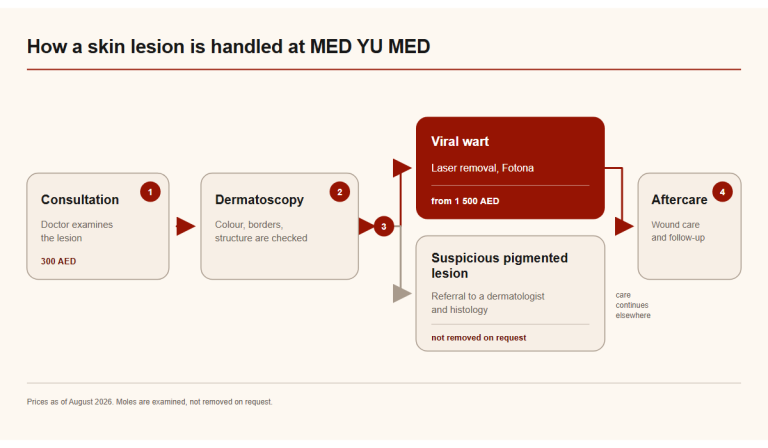
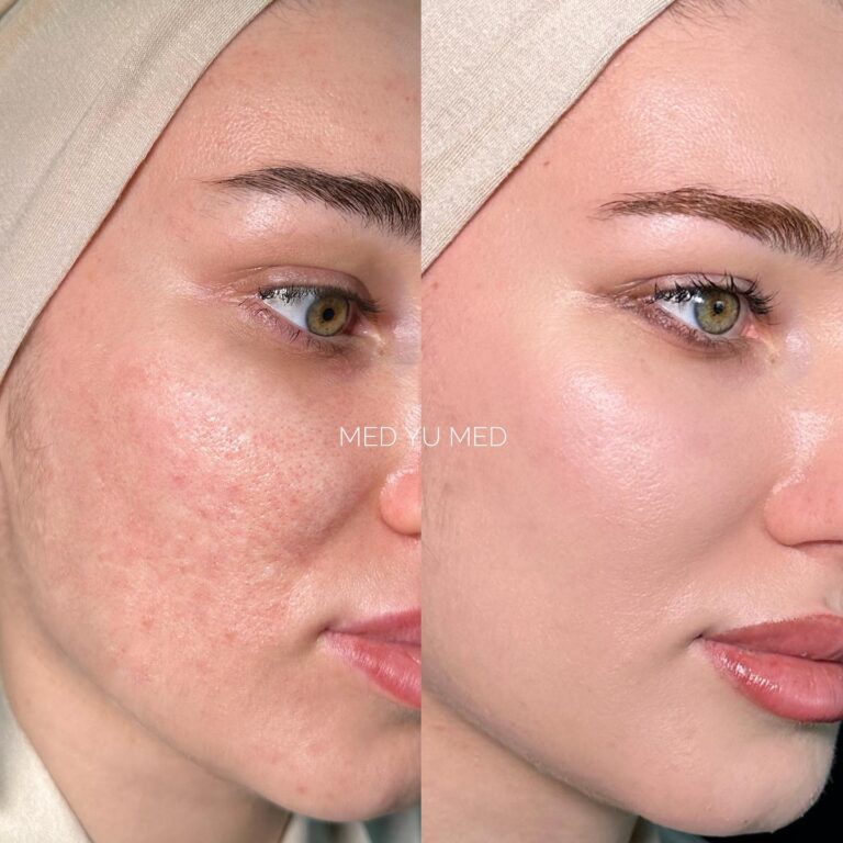
MED YU MED provides the treatments linked below commercially.
Frequently Asked Questions
How do you get rid of milia?
The cyst has to be opened. A doctor makes a tiny incision in the roof with a sterile needle or blade and lifts out the keratin core with an extractor or curette. Widespread lesions may be treated with topical retinoids, electrodesiccation or cryotherapy instead. Scrubs and acne washes act on open follicles and leave a sealed cyst where it was.
Why can’t I squeeze milia out?
A milium is a closed sac with no opening to the surface, unlike a blackhead. Pressure has nowhere to push the keratin, so the bump returns and the skin above it bruises. On eyelid skin, squeezing risks broken capillaries and marks that last longer than the milium would have.
Do milia go away on their own?
Newborn milia do, within a few weeks of birth, and they affect 40% to 50% of healthy full-term babies. In children and adults, StatPearls notes that the other forms of primary milia and secondary milia may not resolve on their own.
What causes milia in adults?
For ordinary milia around the eyes, often nothing identifiable. Documented causes cover skin that healed after burns, dermabrasion or blistering rashes, and topical medications including hydroquinone, 5-fluorouracil and corticosteroids. Cumulative sun exposure produces a related entity called colloid milia on the cheeks and temples.
Is it milia or something else?
Sebaceous hyperplasia, comedonal acne, flat warts, xanthelasma and syringomas all get mistaken for milia, and milia-like cysts appear on dermoscopy in seborrhoeic keratoses and in some basal cell carcinomas. Dermoscopy usually settles it, and where it does not, a biopsy does. Any bump that grows, bleeds or changes colour should be examined rather than extracted.
This article was reviewed by the MED YU MED medical team (DHA-licensed physicians). Last reviewed: 3 August 2026.
Medical disclaimer: This page is for informational purposes only and does not replace a medical consultation.
License statement: MED YU MED operates under DHA License 6589480.
Individual results caveat: Results vary by diagnosis, skin type, and individual response.
Medically reviewed by